Understanding the physical laws governing viruses could lead to the development of new treatments
Read more
Super Abril
super.abril.com.br

Understanding the physical laws governing viruses could lead to the development of new treatments

Viruses obey the laws of physics, and understanding these principles opens the path to creating new therapeutic approaches. This text, written in collaboration with Karin Wuertz-Kozak and Maureen Ferran, was originally published on The Conversation website, which collects materials from researchers.

The attraction of biomedical engineers and biologists to mechanobiology—the study of how physical forces affect biology—is partly due to the beauty of physics at the cellular level. Physics is present everywhere: from how artery wall cells react to blood flow, to how vertebral cells support weight, or how lung cells cyclically expand with each breath.

Since every structural element of the body is subjected to specific combinations of physical forces, the viruses that infect it must also interact with these forces. The way viruses interact with these forces influences their ability to infect cells and cause serious diseases.

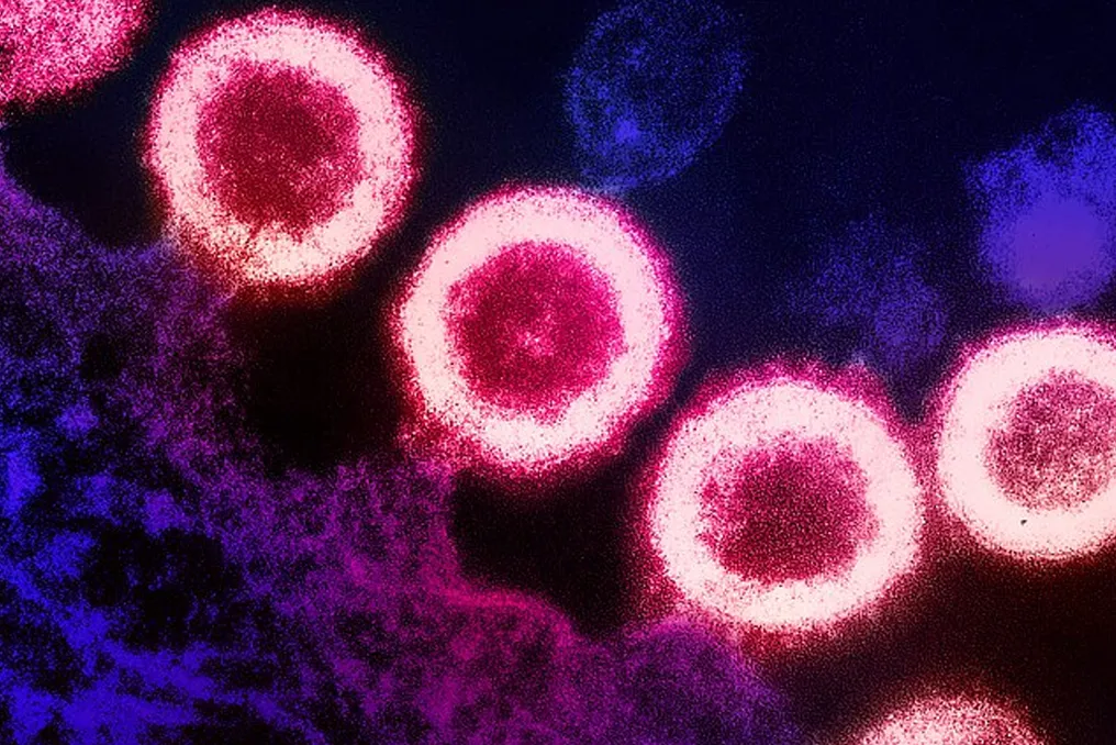
When microscopic viral particles enter the body, they depend on the mechanism of host cells to continue their life cycle. Many viruses first bind to receptors on the cell surface. The movement of the cell membrane, provided by the internal cytoskeleton, causes it to curve and engulf the invasive particle, forming a tiny vesicle that transports it inside. This process, called endocytosis, leads to the cell absorbing the virus.

After entering, the virus moves through the internal structure of the cell, releases its genetic material, and uses the cellular machinery to produce copies of itself. Then, new viral particles leave the cell to infect neighboring cells.

Each of these stages is inherently physical. The tension of the cell membrane can promote or hinder viral incorporation. The cytoskeleton, which transports these viral particles, is the same structure that cells use to sense and apply pressure to the environment. Furthermore, the stiffness of the cellular environment determines how the cell spreads and organizes, which can alter its susceptibility to the invader.

A healthy person usually recovers quickly from an infection. However, people with pre-existing conditions, such as pulmonary fibrosis—where lung tissue hardens and can no longer fully expand—may have a viral infection worsen their condition. This mechanical predisposition of tissues to infection is one reason healthcare workers advise avoiding contact with the elderly and immunocompromised during illness.

Our lab investigates the mechanobiology of different tissue types to understand how everyday physical stresses can exacerbate diseases. For example, repeatedly lifting and carrying heavy objects can wear down intervertebral discs, and mechanical ventilation can overstretch and damage the lungs.

However, culturing cells on a plastic Petri dish—a standard method in most virology labs—only shows part of the picture. The plastic used for tissue culture is significantly stiffer than living tissue, resembling a working surface more than a lung. Cells perceive this difference during experiments; the stiffness of the environment can influence both their behavior and their response to infection.

To bring the mechanics of the human body into the lab, we culture lung cells on thin layers of a flexible silicone polymer called PDMS, which we tune to match the softness of real lung tissue. These layers are placed in small chambers that cyclically stretch and relax upon computer command, which dictates the degree and duration of stretching. This allows us to simulate the physical movements of breathing.

We can use a genetically modified virus to make infected cells glow red under a microscope, tracking how it spreads between cells. But how do we track changes in cell mechanics during infection? Our team monitors the activity of two important proteins called YAP and TAZ, which act as cell force sensors. When tissue experiences stiffness or tension, these sensors move into the nucleus and activate genes related to growth and repair. In the absence of such forces, as typically occurs in softer tissues, YAP and TAZ remain inactive and outside the nucleus.

It is known that some viruses utilize this same signaling pathway; therefore, studying YAP and TAZ can provide insight into how mechanical forces and infection influence each other. Using the CRISPR gene editing tool, we can attach a fluorescent marker to YAP or TAZ and observe their activation in real time under a microscope as they respond not only to mechanical forces but potentially also to infection.

With this platform, we can better answer questions such as: will lung cells respond differently to infection under various forces? Or will these forces change the efficiency of viral infection? Currently, our preliminary results show that the stiffness of the cellular environment affects the degree of cell infection: lung cells grown on stiffer surfaces, similar to fibrosis, appear to be more heavily infected.

Understanding the mechanics of viral infection is of particular interest to researchers and physicians, given the constant prevalence of respiratory infections worldwide. The common cold, COVID-19, and influenza are just a small fraction of viral infections: it is known that over 200 types of viruses infect humans.

Mechanobiology may be as important for the infection cycle as its biochemical or immunological aspects. Under the microscope, the differences between stiff and congested lung tissue and soft, healthy lung tissue are evident, and viruses can move adaptively in these environments.

As scientists map these mechanical signals, new opportunities emerge for preventing and treating infections. Among these are therapies that can reduce physical stress in tissues, or even biomarkers that can inform patients whether they are susceptible to tissue hardening or certain infection pathways and the potential damage they might cause. The human body is as mechanical as it is chemical, and this applies to the infections it encounters.


Similar stories

Shamond virus, previously thought to affect animals, infects and kills human in France
Read more
super.abril.com.br

Shamond virus, previously thought to affect animals, infects and kills human in France

According to European health authorities, a person with a weakened immune system was infected with the Shamond virus and died as a result of this infection in France. This case is significant because it was previously believed that the pathogen exclusively affects animals, particularly cattle, and this is the first registered case among humans.

Despite this incident, experts and health authorities note that the probability of an outbreak or epidemic of this virus remains low.

The Shamond virus was discovered in Nigeria in 1965. It primarily infects ruminant animals such as cows, sheep, and goats, causing fever, diarrhea, and reduced milk yields. In adult animals, it is rarely fatal.

Transmission of the virus occurs through bites of mosquitoes of the genus Culicoides, known as maruvi or powder mosquitoes. This virus is a relative of the Oropuche virus, which has affected thousands of Brazilians in recent years.

In July 2026, an outbreak caused by two new Shamond variants was detected in livestock in Europe, initially in Switzerland and Germany. The disease spread to farms in France, Spain, Portugal, and other countries.

The man's death occurred on October 9th. He was over 60 years old, lived in a rural area of the Rhône-Alpes region, where the virus was already circulating among animals, and was battling cancer. The symptoms he exhibited included diarrhea, vision loss, confusion, and other neurological problems. His condition progressed to encephalitis, which proved fatal.

According to a report published in the journal Eurosurveillance, it is most likely that the man was infected after a maruvi bite. Less likely hypotheses include infection through consuming contaminated food or after blood transfusion.

The patient suffered from severe leukemia and had a weakened immune system, which, according to scientists, likely exacerbated his condition. A weak immune system can allow the virus to remain in the body longer and reach more organs. Autopsy revealed high viral levels in the man's brain, liver, and spleen.

The Shamond virus is not detected by standard blood tests; doctors had to sequence the RNA present in the patient's samples. The virus genome found in the man's body was very similar to that circulating in animals, with an emphasis on five mutations. Researchers do not yet know if these changes make the virus more transmissible to humans.

Artificial intelligence is capable of creating viruses that infect internet-connected household appliances
Read more
olhardigital.com.br

Artificial intelligence is capable of creating viruses that infect internet-connected household appliances

With the development of artificial intelligence (AI), devices connected to the internet and exchanging data in the so-called 'Internet of Things' (IoT) can become a source of problems. According to a report by Anthropic published in October, AI-based malware can be installed on such devices and spread autonomously.

Computer viruses that move independently from one machine to another are known as 'worms'. They follow a predefined plan, utilizing known security vulnerabilities or poorly protected access points. Typically, solving this issue involves fixing bugs and closing gaps in the security system. However, in the case of AI-based 'worms', the situation becomes significantly more complex and dangerous.

This is because this more advanced type of 'worm' is capable of independently solving problems and adapting to different types of devices. Researchers at Anthropic, the developer of Claude, demonstrated that AI applications can function without the need for a large data center.

In a controlled test environment, researchers created simulations of eight Android devices. The tested AI model was installed on one of these devices, then copied from it and infected the others. In four out of five attempts, the model reached all eight targets, and in one, it infected seven.

If such an AI model were combined with malicious software, the 'worm' could learn to bypass security measures. According to Anthropic developers, this would take approximately one to two days. Furthermore, the AI could potentially connect infected devices into a botnet.

Although an infected device has limited computational capabilities, the more devices that are infected, the faster the 'worm' can spread. Connected devices can theoretically be disconnected from the network, and security vulnerabilities can be patched. Nevertheless, the decentralized structure of such malware will greatly complicate its localization.

Anthropic describes this type of infection as 'digital slime mold,' which grows slowly. The company believes that smaller AI models will be able to achieve this level of propagation capability as early as 2026.

Brazilian scientists investigate the use of sound waves to inactivate Covid-19 and Influenza A viruses
Read more
super.abril.com.br

Brazilian scientists investigate the use of sound waves to inactivate Covid-19 and Influenza A viruses

In the context of combating the Covid-19 pandemic, although vaccines and medications have been crucial, the possibility of new emergencies stimulates the continuation of scientific research. An initiative involving researchers from Unesp and USP is exploring the potential of sound waves to destroy microorganisms.

The findings were published in the scientific journal Scientific Reports. While vaccines and drugs act through chemical and biological pathways, the recent investigation focuses on therapies based on physical properties, such as using ultrasound to rupture the structure of respiratory viruses and control viral load.

In in vitro experiments, Brazilian scientists demonstrated that ultrasound waves managed to destabilize both the Covid-19 virus (SARS-CoV-2) and Influenza A (H1N1). This process led to the rupture of the viral envelope, fragmentation of particles, and a decrease in these viruses' ability to infect cells.

The application of these physical techniques in human treatment presents challenges, as many in vitro or environmental disinfection methods require high intensities, making them unsuitable for living tissues. For example, ultraviolet radiation inactivates viruses quickly but also carries a high risk of tissue damage, similar to other forms of non-ionizing radiation.

In preliminary tests, researchers noted that exposure to ultrasound was effective without registering significant changes in the temperature or pH of the samples. This paves the way for a purely physical viral inactivation strategy, which utilizes the mechanical characteristics of the particles, such as their shape, instead of relying on chemical agents or high doses of radiation.

The multidisciplinary team includes specialists in physics, virology, and materials characterization. Members include Professor Odemir M. Bruno from the Physics Institute at USP in São Carlos; Professor Carlos José Leopoldo Constantino and postdoctoral fellow Gilia Cristine Marques Ruiz, both from the Faculty of Science and Technology at Unesp, on the Presidente Prudente campus.

The investigation began during the Covid-19 pandemic, when Bruno's group was developing sensors for viral detection. It was during this period that a theoretical model emerged indicating that viral particles could resonate with certain acoustic waves, which spurred the research starting in 2021.

The central concept is resonance. When a sound wave encounters an object capable of vibrating at a corresponding frequency, the transferred energy can accumulate and amplify the oscillation, potentially leading to the collapse of the object. Bruno illustrates this by comparing it to breaking a crystal glass with a violin, emphasizing that the ultrasound approach uses a gentle and harmonic strategy.

The spherical shape of the virus is crucial because it allows the vibration to distribute uniformly throughout the structure, optimizing the resonance effect. As it approaches the particle's natural vibration frequency, ultrasound transfers energy more efficiently, generating repeated deformations in the viral envelope. If the vibration is intense enough, the mechanical stresses overcome the structural resistance, leading to deformation, rupture, and particle fragmentation.

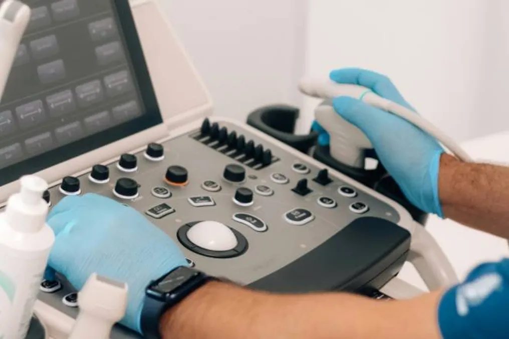
This morphological change results in viral inactivation, causing the virus to lose its ability to replicate and infect. The team employed high-frequency ultrasound, in the range of 3 to 20 megahertz (MHz), using diagnostic imaging equipment. In one specific test, 7.5 MHz was applied for five minutes to analyze structural changes.

To rule out heating or chemical changes, researchers monitored temperature and pH, finding that both remained stable under the resonance conditions. This data is vital because it distinguishes the studied mechanism from cavitation, an ultrasound effect at lower frequencies (kilohertz) that generates heat and shockwaves, useful only in asepsis, but not in therapy.

Bruno compares the ultrasound used to that used in prenatal exams, describing it as harmless. However, he emphasizes that therapeutic application still requires more study and understanding of the phenomenon before advancing to live subject testing.

Techniques such as scanning electron microscopy and atomic force microscopy revealed changes on the surface of SARS-CoV-2: spherical particles began to have irregular contours, smaller diameters, and envelope ruptures. There was also a reduction in the average diameter; while untreated SARS-CoV-2 samples had about 107 nanometers, smaller components appeared after ultrasound.

In the case of H1N1, the effect was even more drastic, with no sign of particles detected in the analyzed size range. At this point, the collaboration of Unesp in Presidente Prudente was fundamental for characterizing nanometric particles, which are difficult to observe with conventional optical microscopy.

Gilia explained that the team used Dynamic Light Scattering (DLS). This method measures the average size of particles dispersed in a liquid by analyzing the fluctuations of light scattered by them. DLS allowed measuring the sizes of virus samples before and after exposure to ultrasound, confirming the fragmentation of SARS-CoV-2 and the absence of H1N1.

Adapting DLS for real viral samples required equipment optimization, algorithm adjustment, and attention to factors such as solvent and thawing.

In addition to physical destruction, scientists evaluated the biological impacts. Treated and untreated SARS-CoV-2 samples were used to infect Vero E6 cells. After 24 hours, analysis of viral replication showed a reduction in infection after ultrasound exposure, with the strongest effect near 7.5 MHz.

The study also included the Gamma and Delta variants. Although ultrasound reduced the infectivity of these variants, they showed greater resistance, and the effectiveness varied depending on the frequency and equipment used.

Bruno points out that the ideal resonance frequency may depend on the physical characteristics of the virus (size, geometry, and mechanical properties), and not directly on its genetic sequence. He states that, in principle, spherical viruses could respond to specific frequency ranges, opening a pathway less dependent on molecular mutations.

However, the differences observed in the Gamma and Delta variants suggest that physical characteristics such as rigidity, envelope thickness, and viscoelastic properties may alter the modes of vibration and, consequently, the efficiency of resonance.

Popular