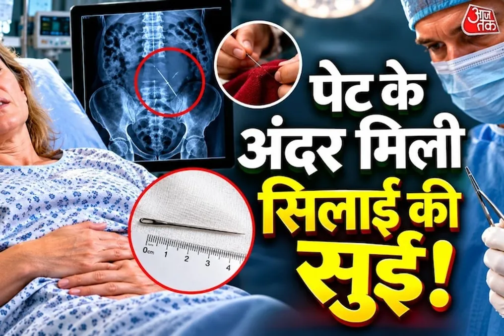
Sewing needle found in woman's abdomen; doctors were surprised after hospital visit
Read more
Aaj Tak
www.aajtak.in

Sewing needle found in woman's abdomen; doctors were surprised after hospital visit

Sometimes minor health issues can have unexpected causes. This happened to a 50-year-old woman in Australia. She suddenly experienced severe abdominal pain. When she sought medical attention at the hospital, doctors were shocked: a sewing needle, about 4 centimeters long, was discovered inside her abdomen. The woman herself did not know how this object had entered her body.

This case was documented by doctors from John Hunter Hospital in Australia in a medical report. According to the report, the woman felt intense abdominal pain that lasted for about a day. Subsequently, she went to the emergency department. Upon examination, mild tenderness was noted upon palpation of the abdomen, along with a rapid pulse—heart rate reached approximately 130 beats per minute. General blood tests showed no serious abnormalities. Doctors then performed a computed tomography (CT) scan, which revealed a metallic object about 4 centimeters long in the left side of the abdomen. Initially, the doctors could not determine how this object had gotten inside.

The woman was kept under observation all night. When the pain subsided, she was discharged with a warning to return to the hospital if the pain recurred. However, the pain returned the next day. An X-ray revealed that the metallic object inside the abdomen had shifted. At this point, the woman recalled something old: before the incident, she had been sewing and had fallen asleep. She hypothesized that she might have accidentally lain on a sewing needle in her sleep, allowing it to penetrate her skin without her realizing it.

The main concern for the doctors was that the moving needle could damage the intestines or blood vessels. This could lead to serious internal injuries, bleeding, or infection. Therefore, the doctors decided to perform surgery to remove the needle using laparoscopic surgery, which allows intervention through small incisions without the need for large cuts.

During the operation, doctors found a small collection of blood near the tissue supporting the small intestine. Locating the needle itself was difficult. Using several X-rays and specialized equipment, they were able to pinpoint the needle's location. It was determined that the needle was lodged in a specific fatty tissue inside the abdomen called the Greater Omentum, which is a layer of fat around the abdominal organs. The needle was near a part of the large intestine. The doctors carefully extracted it. Fortunately, during the operation, the woman's intestines and other organs were unharmed.

The day after the successful removal of the needle, the woman remained under observation. She was discharged the day after the surgery. Later, she developed a mild infection around her navel, which the doctors treated with oral antibiotics. In their report, the doctors noted that foreign objects entering the body can change position, posing a risk of intestinal perforation, infection spread, and blood vessel damage.


Similar stories

Woman finds 40 dead bedbugs on hotel bed in Britain; has to seek hospital care
Read more
www.aajtak.in

Woman finds 40 dead bedbugs on hotel bed in Britain; has to seek hospital care

A guest staying at a hotel for work encountered an unpleasant situation that prompted her to thoroughly check the bed before sleeping. Although people expect cleanliness and comfort in hotels, something happened to this woman that makes one think about carefully inspecting beds.

The first night passed normally, but the next morning the woman noticed red spots on her body. Initially, she did not pay it any mind, but after removing the sheet and duvet from the bed, she was shocked. About 40 dead bedbugs were found on the bed.

This incident occurred in the city of Exeter, Britain. 46-year-old Nicola Jenkins, who lives there, stayed at the Holiday Inn Express Exeter East for work. She booked a four-night stay, arriving at the hotel on October 12. She did not expect her hotel stay to turn into such a negative experience.

According to Nicola, there were no problems on the first night. However, the next morning when she woke up, she felt itchy all over her body. After showering, she saw red marks on her face, neck, arms, back, and abdomen. At first, she assumed they were insect bites, but the number of marks kept increasing. Since Nicola had previously encountered bedbug bites, she suspected there might be bedbugs in the bed.

After this, Nicola decided to check her bed. When she removed the duvet, she saw a crawling bedbug and took a video. A thorough inspection of the sheets and bed revealed about 40 dead bedbugs. Nicola reported that she had over a hundred places on her body affected by bedbug bites, including red marks on her forehead, cheeks, neck, waist, and arms. Seeing such a large number of bedbugs, she panicked.

Following this incident, Nicola complained to the hotel reception desk. In response, the hotel provided her with another room. Nevertheless, Nicola noted that she was informed that since her office made the payment, a direct refund was not possible. During this incident, she was also offered a free cup of coffee.

However, Nicola's problems did not disappear. She decided to leave the hotel and stay elsewhere. Despite this, the itching and redness on her body continued to worsen.

Nicola recounted that after the bedbug bites, her skin began to itch and burn severely, creating a burning sensation. As the situation worsened, she visited a pharmacist who prescribed antihistamines to relieve the itching. But her condition did not significantly improve. On October 19, her left arm became severely swollen and completely red, after which she had to go to the hospital's Accident & Emergency (A&E) department. After examination, doctors prescribed her a course of steroids.

Nicola shared that this incident caused her a lot of inconvenience; she could not attend an event like a children's party on the weekend and had to stay home. After treatment, her condition gradually improved.

Following the incident, Nicola's office also filed a complaint against the hotel. The hotel contacted her office and acknowledged the problem related to bedbugs. The hotel offered Nicola free accommodation for the weekend at another hotel. However, Nicola declined this offer, stating that after such an experience, she would never want to stay in this hotel chain again.

A representative of Holiday Inn Express Exeter East stated that the hotel takes guest cleanliness and safety seriously. He reported that he received a complaint regarding this incident and is conducting an investigation. The hotel also noted that they are in contact with the woman. This case has once again raised questions about room cleanliness and bed inspection in hotels.

Stone structure formed from fetus skeleton found in abdomen three years after pregnancy
Read more
www.aajtak.in

Stone structure formed from fetus skeleton found in abdomen three years after pregnancy

Cases related to medical science that cause astonishment among people periodically appear in the world. One such unusual case occurred in Somaliland. There, a 42-year-old woman had a stillborn fetus in her abdomen for approximately three years. Notably, over time, this fetus turned into a hard, stone-like structure. After examination, doctors removed it. In medicine, this rare condition is called 'Lithopedion' or 'stone baby.'

The woman felt pain and heaviness in the lower part of her abdomen for the last three years. Initially, she considered it a normal problem, but the pain gradually intensified. When she sought medical attention for an examination, the truth that emerged shocked everyone.

According to medical reports, about three years ago, the woman developed symptoms similar to pregnancy. During that period, her abdomen began to enlarge, and she felt movements resembling those of a baby. However, these sensations soon stopped, and her abdomen began to slowly decrease. The woman did not undergo hospital examinations or consult with doctors during this period, unaware of what was happening in her body. For the next three years, she experienced mild pain and a feeling of heaviness in her lower abdomen, which became more pronounced in recent months. Eventually, she was admitted to the hospital for treatment.

During the hospital examination, doctors felt a hard and unusual lump in her abdomen, which extended almost to her navel. Blood tests showed the woman had mild anemia. Furthermore, a pregnancy test was negative. An ultrasound was then performed, which showed a hollow uterus, but a hard, calcium-composed structure was visible in the abdomen. To better understand the situation, doctors performed a CT scan. On the resulting 3D images, they saw the skeleton of a fetus inside her abdomen, clearly showing the skull, spine, ribs, and bones of the arms and legs. The size of this entire structure was about 18x12x10 centimeters. After the examination, doctors identified it as lithopedion, or a stone baby.

Although the term 'stone baby' sounds like a movie plot, it is an extremely rare phenomenon related to medical science. To understand this, one must first consider a normal pregnancy. Usually, after conception, the baby develops in the mother's uterus. However, in some cases, the fetus begins to develop not in the uterus, but directly in the abdominal cavity. This condition is called abdominal pregnancy, or ectopic pregnancy in the abdomen. In such cases, normal fetal development is hindered. Often, the fetus dies after some time. After this, the woman's body begins to slowly try to expel or absorb the dead fetus. But if the fetus is large, the body cannot fully absorb it. In this situation, the body begins to form a hard layer of calcium around the fetus. Over time, calcium accumulates in this layer, and the fetus turns into a stone structure. This is why it is called a 'stone baby.'

Essentially, the body does this for self-protection. The formation of the calcium layer reduces the contact between the tissues of the dead fetus and surrounding organs, thereby reducing the risk of damage. Nevertheless, the presence of such a structure in the abdomen can cause the woman pain, abdominal heaviness, and other health problems. Sometimes this is discovered years later when an X-ray or scan is done while examining another problem.

After the situation was fully understood through examinations, doctors decided to perform surgery. During the surgical procedure, doctors removed the fetus covered with a calcium layer from the woman's abdomen. The day after the operation, the woman experienced a slight fever and nausea, but these discomforts passed after treatment. She was then discharged from the hospital. Doctors monitored the woman's condition for the following six months. During this time, her condition improved, and the abdominal pain completely disappeared.

The rarity of this disease can be assessed by the fact that only 330–340 such cases have been registered in medical records over the last approximately 400 years. Typically, such cases are discovered when women undergo examinations due to persistent abdominal pain or other problems. In some women, this condition persists for decades, and they may not even know about it. In this case, the woman was only 42 years old, and the fetus had been in her abdomen for about three years. The medical community emphasized the importance of timely examination and medical care during pregnancy. This case demonstrates that any unusual symptoms related to pregnancy should not be ignored; a timely visit to a doctor allows serious problems to be detected.

Popular