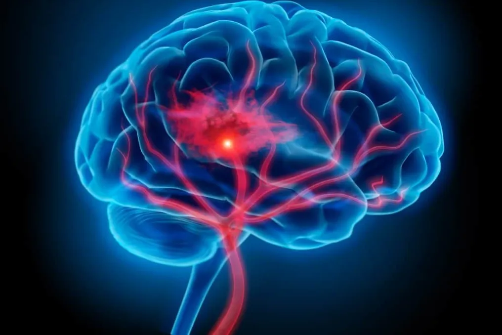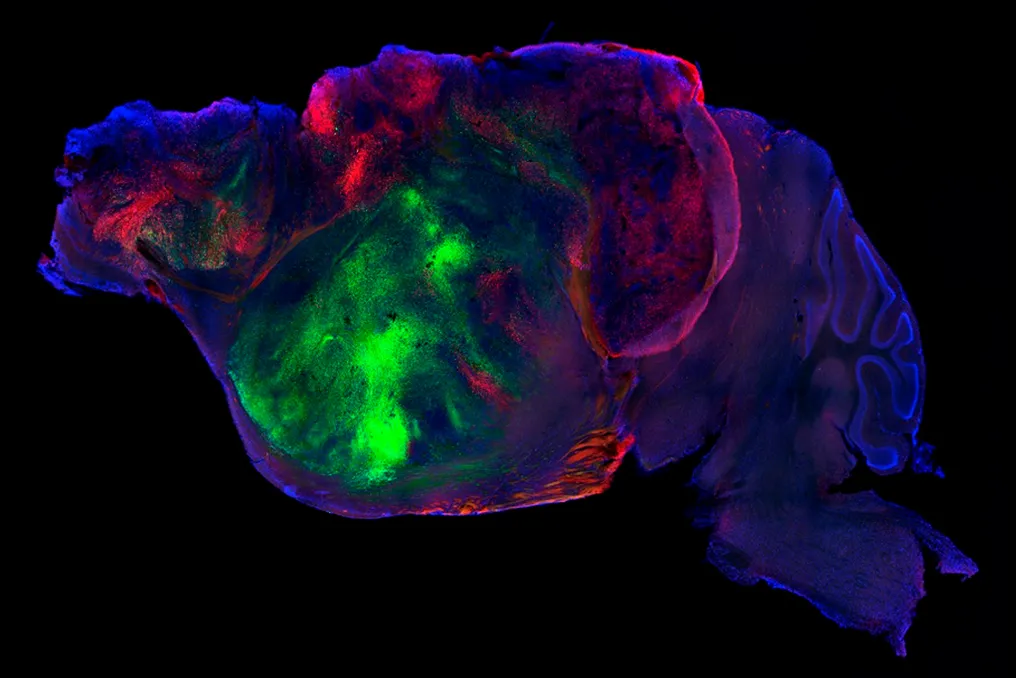
At Stanford University in the United States, a team of scientists is monitoring about twenty mice that, while appearing normal at first glance, contain an extraordinary element in their brains: millions of human neurons.
A new study published in the journal Nature describes how neuroscientists transplanted human brain cells into the brains of mice, filling incomplete areas of the nervous system. This procedure creates a new experimental model that may help identify causes or develop treatments for neuropsychiatric disorders, such as certain types of dementia or cerebral palsy.
This work marks a significant advance in efforts to recreate the human brain outside the body. Analyzing living neurons has been a persistent challenge in this field of research for several decades.
An alternative emerged in the early 2010s with the development of structures called organoids. Instead of examining a human organ directly, researchers began to be able to culture smaller, simplified versions of body tissues in the laboratory. For example, from skin cells, various cerebral organoids can be generated that mimic certain functions and characteristics of a real brain.
However, these replicas have limitations, as they lack blood vessels and are very small. Sergiu Pasca, one of the researchers responsible for the discovery of organoids and the current coordinator of the mouse study, stated in a release that it would be impossible to study complex human behaviors within a cell culture plate.
The attempt to overcome this limitation occurred in 2018, when scientists implanted cerebral organoids into the brains of mice. Subsequently, in a 2022 study, these cells were transplanted into the visual center of the animals, demonstrating that both mouse and human neurons responded to light.
More recently, in 2024, another study led by Pasca investigated Timothy syndrome, a rare genetic condition associated with epilepsy and severe autism, using organoids in young mice. This method allowed the team to assess the effectiveness of antisense oligonucleotides, a specific type of drug, against the disorder.
Although similar to the most recent study, the 2024 experiment used a more basic transplant, inserting human brain cells next to the brain tissues of young rodents, which were already rapidly developing, forcing the human organoids to compete for space.
In the current experiment, researchers used genetically modified mice that do not develop the cortex—the outer layer of the brain responsible for cognition, memory, and decision-making, and which is disproportionately larger in humans to support language and complex thought. In this location, inside the cranial cavity, there was an empty space filled with cerebrospinal fluid.
The surgery to inject the human cells was performed a few days after the birth of the mice, when primary brain connections were already established. Into this absent cortical area, hundreds of thousands of human cells were introduced. Without competition, they multiplied rapidly; in two or three months, the quantity almost quintupled, reaching about 4 million and occupying more than 90% of the space previously occupied by fluid.
These human cells, still immature (comparable to those of a fetus in the third trimester of pregnancy), activated and formed connections as the mouse lived. Furthermore, the human tissue managed to generate a specialized type of neuron linked to social cognition, fusiform neurons, which had never been created in a laboratory. These neurons are the first cells to die in cases of frontotemporal dementia.
The modified mice showed no visible differences compared to common mice, both in their movements and in performance on cognitive and memory tasks. However, an exception was observed when subjecting both types of mice to low oxygen conditions. While this deprivation can cause severe damage to the cortex in human infants, leading to cerebral palsy, the same does not occur in mice; however, the mice with human brain cells exhibited symptoms analogous to those of individuals with cerebral palsy.