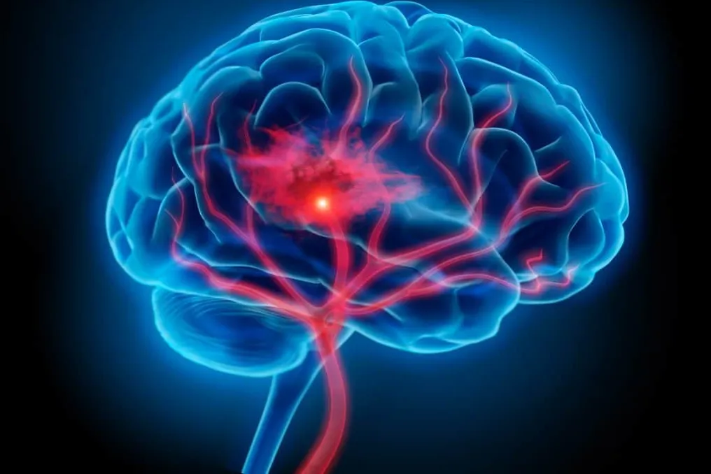
Researchers at Stanford Medicine have developed a new methodology that allows for the cultivation of human brain tissue inside genetically modified mice. These animals are engineered not to develop most of their cortex and hippocampus. The implanted graft demonstrated the ability to grow, form blood vessels, and establish functional connections with the rodents' nervous system.
These so-called 'xenocortical mice' offer a valuable tool for investigating brain development and various neurological pathologies. Furthermore, the experiment allowed for the observation of types of human neurons that are difficult to replicate in traditional laboratory settings, enabling testing of this tissue's reaction to changes associated with different health conditions.
To conduct the study, scientists genetically altered the mice, preventing extensive development of the cortex and hippocampus. Some animals served as controls, remaining without the graft, while others received human cortical organoids shortly after birth.
As the animals grew, the human tissue occupied the available space. Over a three-month period, this tissue increased its initial volume by almost five times, accounting for over 90% of the cortical volume. The graft also became electrically active and developed blood vessels, sending projections to the spinal cord.
Sergiu Pasca, a professor at Stanford and author of the study, clarified that although the animals possess a mouse nervous system, sensory organs, and subcortical structures, the notable aspect is that most of the cortical tissue is of human origin, and the human neurons are able to grow, integrate, and create functional connections with the rest of the mouse's nervous system.
It is important to note that the transplanted tissue has not yet reached the maturity of a human cortex. Even after approximately six months, it remained immature and lacked the typical organized layers of an adult cortex. However, researchers identified neurons that showed difficulty in being produced in other models, including cells similar to von Economo neurons, which are elongated structures found in specific areas of the human brain.
Among the potential applications of this model is the study of memory. In a test conducted with a Y-maze, the xenocortical mice demonstrated performance better than expected by chance. Animals lacking cortex and hippocampus showed difficulties in remembering which leg of the maze they had visited. However, Pasca warned that this result does not allow one to state that human neurons are directly responsible for the observed behavior.
The incorporation of human brain tissue also raises important ethical dilemmas. The team included Stanford bioethicists and an external committee composed of neuroscientists, ethics specialists, legal experts, and patient representatives. The animals were rigorously monitored throughout the study to detect any unforeseen biological or behavioral effects.
Another central concern lies in determining whether the progressive insertion of increasingly complex human neural tissue into an animal's nervous system can generate emergent or novel properties that require additional ethical consideration. The study, published in the journal Nature, positions the xenocortical mice as a tool to examine the human brain in a living organism, while maintaining the challenge of distinguishing the effects of human cells from those generated by the animal's nervous system.