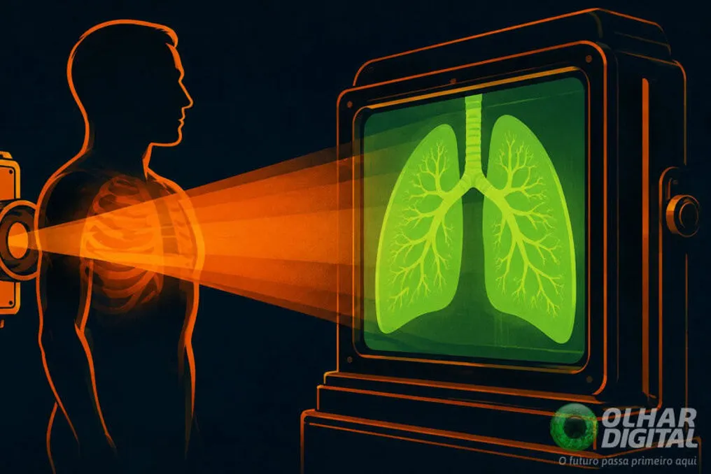
You may have heard or read that the X-ray beam was invented by a Brazilian. However, this is not entirely accurate. Nevertheless, the doctor Manoel Dias de Abreu deserves his place in the history of radiology. Although he may not have invented the technology itself (which belongs to the German physicist Wilhelm Conrad Röntgen), he developed an improvement to the X-ray beam—abrography.
Both methods use X-ray radiation to view the internal contents of the human body, but they differ in operating principle and cost.
It should also be noted that thanks to abrography, the Brazilian doctor spearheaded a revolution in public health in the country.
The history of radiology began on November 8, 1895, in Röntgen's laboratory. During experiments with electricity in nearly airless glass tubes, the physicist noticed that nearby paper began to glow. He discovered an 'invisible ray' emanating from the tube that passed through solid objects.
Since Röntgen did not know exactly what this radiation was, he temporarily called it the 'X-ray beam.' In mathematics, the letter 'X' usually denotes an unknown quantity.
The physicist then established that these rays pass through skin and muscles but stop at dense materials, such as bones. On December 22, 1895, he asked his wife, Anna Bertha Röntgen, to place her left hand on a photographic plate. The scientist then turned on the apparatus and allowed the radiation to act for about 15 minutes.
Upon developing the film, Röntgen saw an image that entered history: the silhouette of his wife's hand bones and the outline of the wedding ring she was wearing at the time of the exposure. This was the first X-ray image in the world. In 1901, the scientist received the Nobel Prize in Physics for his discovery.
Going back a bit, the first X-ray image in Brazil was taken in 1896. At that time, it was a revolutionary technology, but it was very lengthy and expensive. Mass use among the population was impractical, yet necessary.
At that time, the world was facing a tuberculosis epidemic—a slow-developing bacterial disease that people discovered when it was already too late for treatment. In capitals such as Rio de Janeiro (RJ) and São Paulo (SP), the situation was catastrophic.
The question arose: what if mass, fast, and inexpensive X-ray images could be taken? This was the question pondered by the doctor from São Paulo, Manoel Dias de Abreu. His idea was to combine radiology with ordinary photography.
This device did not work immediately. Abreu tried to create this system in Paris in 1919, but unsuccessfully. Then, in 1924, he repeated the attempt, and it failed again. The problem was that the X-ray screens of that time glowed weakly, which resulted in very dark photographs.
Only in 1936, still in Rio, was Abreu able to obtain his first clear images thanks to the technology of brighter screens. Initially, the doctor named the method X-radiography in honor of the physicist Wilhelm Conrad Röntgen. Later, in 1939, the method became known as abrography in honor of the doctor.
Being one hundred times cheaper than conventional X-rays, abrography allowed millions of people to be diagnosed with tuberculosis before it was too late. For example, the disease could be detected even before any symptoms appeared.
Casa Lohner, a subsidiary of Siemens, manufactured the first abrography devices in Rio de Janeiro based on the doctor's drawings and instructions. Thus, his method quickly spread worldwide, significantly aiding the fight against tuberculosis.
Significant advancements in radiology brought Abreu several nominations for the Nobel Prize, but the doctor never received it.