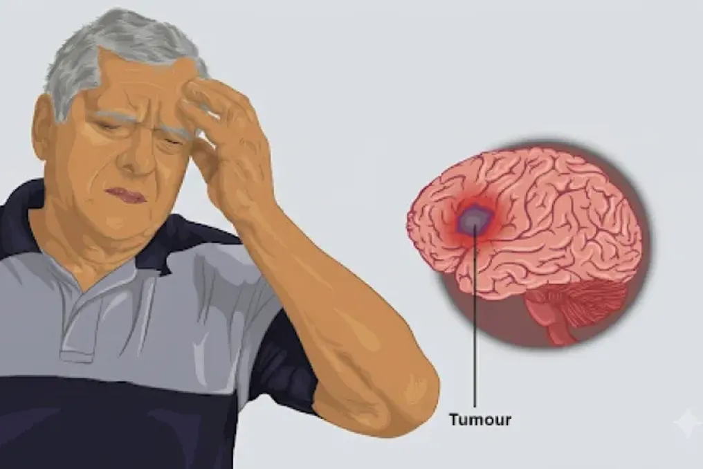
Glioblastoma is one of the most aggressive forms of brain cancer because it spreads rapidly, is resistant to treatment, and is difficult to surgically remove. Even after surgery, radiation therapy, and chemotherapy, patients usually live only from six months to a year, and the tumor almost always returns, becoming more fast-growing, treatment-resistant, and prone to spreading into surrounding brain tissues.
A series of studies conducted in the mechanobiology laboratory of Professor Abhijit Majumder at the Indian Institute of Technology Bombay (IIT Bombay) suggests that part of the problem lies in the methods used to study these tumors. The results provided an explanation for the aggressive, recurrent nature that goes beyond genetics.
Scientists discovered that the physical softness of the tissues around the tumor can activate genes that promote cancer development and mask potential drug targets that only become visible when studying cells in conditions mimicking real brain tissue.
In the first study, led by Professor Shilpi Dutt from the Centre of Excellence in Cancer Treatment, Research and Education (ACTREC) Tata Memorial Centre (now at the School of Life Sciences, JNU), in collaboration with Majumder's team, researchers recreated brain tumor recurrence in the lab and published their findings in the journal Matrix Biology. To do this, they irradiated brain cancer cells, destroying about 90% of them. The surviving cells were then cultured and allowed to regrow in the lab, simulating tumor recurrence after treatment.
When these recurrent cells were implanted into mouse brains, they behaved more aggressively than the original tumor cells (also called parental cells), which aligns with clinicians' observations in patients. However, when populations of both parental and recurrent cells were grown separately on standard hard plastic culture dishes, both populations showed identical behavior in tumor growth, invasiveness, or treatment resistance.
Professor Majumder noted: 'We suspected the problem lay in the experimental model itself. Although the brain is one of the softest tissues in the body, like jelly, cancer cells are usually studied on hard plastic surfaces because they are convenient, reproducible, and standardized in laboratories.'
To test whether this mismatch masked important biology, researchers cultured tumor cells on polyacrylamide hydrogels designed to match the softness of brain tissue. On these soft gels, recurrent tumor cells acquired an invasive, elongated, fast-moving morphology that was previously invisible when growing the same cells on plastic.
Professor Dutt emphasized: 'The hard surface did not just dampen the aggressive behavior of the cells; it erased the very distinction between parental and recurrent cells that defines clinical recurrence.'
The implications of this discrepancy also affected which drug targets researchers could discover. By comparing the gene expression profiles of brain cancer cells on both surfaces, scientists identified the protein PLEKHA7, which was upregulated only on the brain-mimicking gel. Its levels were also elevated in biopsies of patients with recurrent glioblastoma. It never appeared on plastic. Blocking PLEKHA7 successfully reduced tumor cell survival and spread.
According to Professor Dutt: 'Thus, a real, functionally validated drug target could have simply been missed by using the traditional culture method that most laboratories still rely on.'
Although 'soft versus hard' comparisons have been done by many groups before, Professor Majumder notes that 'the uniqueness lay in comparing parental tumor cells with recurrent cells and demonstrating that their response to substrate stiffness was different—a difference invisible on plastic.'
By investigating the role of physical signals, such as tissue stiffness, in tumor behavior, Majumder's lab began to explore whether certain proteins and RNAs already known as glioblastoma progression drivers could themselves respond to physical signals.
Dr. Arpita Ghosh, first author of two subsequent studies, focused on NEAT1—a long non-coding RNA (lncRNA) gene regulator associated with tumor growth, invasion, treatment resistance, and the migration of tumor cells to new sites. Dr. Arpita stated: 'While proteins responding to physical forces have been widely studied, lncRNA molecules like NEAT1 have never been considered as potential physical signal sensors.'
Working with Professor Mohit Kumar Jolly's group at the Indian Institute of Science in Bengaluru, Majumder's team cultured brain cancer cells on hydrogels of varying stiffness. Their results, presented in the bioRxiv preprint, showed that NEAT1 levels inside the cells increased three to fourfold on soft, brain-like surfaces compared to hard plastic. When the team used RNA interference to reduce NEAT1 levels specifically in cells grown on soft, brain-like gels, many of their aggressive characteristics decreased.
Dr. Arpita noted: 'The ability of cancer cells to self-renew, spread through tissue, and adopt a more motile, shape-changing form diminished. This was essentially one of the most important findings, showing us that NEAT1 can be a mechano-sensitive molecule capable of translating physical signals into changes in tumor behavior.'
In a related study published in Macromolecular Bioscience, researchers investigated another limitation of the standard lab model. Professor Majumder noted: 'A real tumor is a three-dimensional structure, it is a sphere. But so far we have studied everything in two dimensions, including soft gels. The question is, are we missing something?'
To recreate this architecture, researchers grew glioblastoma cells into three-dimensional tumor spheroids, or tumoroids. These miniature tumors allow cells to organize and interact in ways that more closely resemble real cancers. NEAT1 levels were approximately 3.5 times higher in these 3D models than in cells grown on 2D soft gels. Suppressing NEAT1 using RNA interference slowed cell division and reduced tumor growth. Under the microscope, tumoroids with non-suppressed NEAT1 levels had jagged, protruding edges, a characteristic feature of cancer cells detaching to spread. Conversely, tumoroids with suppressed NEAT1 had smooth, compact borders, suggesting that the cancer was less likely to invade surrounding tissues.'
Dr. Arpita concluded: 'Our findings show that the tumor architecture itself can influence the behavior of cancer-related molecules.'
Furthermore, researchers determined whether this phenomenon extends beyond brain cancer. By comparing 2D and 3D cultures of breast, cervical, liver, and lung cancer cell lines, they found a tissue-specific pattern. Both NEAT1 and its associated lncRNA, MALAT1, increased in breast, lung, and brain cancer cells grown in 3D, but differed in cervical and liver cancers. From this data, the researchers emphasize that laboratory models must match the unique physical environment of each type of cancer to accurately reflect disease biology.
According to Majumder, 'what distinguishes this body of work is the link between mechanics, lncRNA biology, and cancer progression. This has never been done before.'
Collectively, these discoveries indicate that the tumor's physical environment can shape its behavior as profoundly as its genetics, and hard plastic dishes may cause researchers to overlook important drug targets.
Professor Majumder concludes: 'Simply by changing the stiffness, we were able to reproduce what was observed clinically, which was impossible to see on plastic dishes. If anyone has a target for an anticancer drug, they should now include this (soft gels and tumoroids) in the initial experiment.' He adds that the cost of missing this step is already evident: approximately nine out of ten cancer drug candidates fail in human trials despite promising early results.