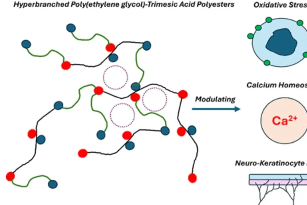
Restoring damaged human tissues requires creating a microenvironment that mimics the complex signaling of the extracellular matrix. Recent advancements in biomaterials have highlighted the critical importance of the relationship between keratinocytes, the primary cells of the outer layer of the skin, and peripheral nerve endings. This communication between neuro-keratinocytes is essential for maintaining skin health and sensitivity.
Development of Novel Biomaterials
Creating a synthetic scaffold that simultaneously supports both cell types and regulates their internal chemistry has long been a significant challenge. A multidisciplinary research group from Calcutta University, National Institute of Science and Education and Research in Bhubaneswar, Lion Elastomers, USA, KIIT Deemed to be University, and the Institute of Elastomer and Tire Research of Hari Shankar Singhania solved this problem. They developed a series of hyperbranched polyesters based on polyethylene glycol and trimellitic acid (PEG:TMA).
Principles of Scaffold Creation
These materials are designed as tunable scaffolds that can be adapted to manage oxidative stress, calcium homeostasis, and the structural needs of regenerating tissue. The researchers used polyethylene glycol as a flexible backbone and trimellitic acid as a trifunctional monomer, which led to the induction of hyperbranching. This process forms a dense network of functional end groups and allows for the regulation of the ratio of soluble, branched components and insoluble, cross-linked structures.
Comparative Analysis of Compositions
By systematically varying the molar ratio of PEG to TMA from 1:0.5 to 1:5, the team obtained five different compositions, designated as S1–S5. The physical properties of these materials varied significantly depending on the TMA content. At lower ratios, the surfaces were smoother and more soluble with minimal cross-linking, whereas higher TMA concentrations yielded denser, stiffer, and more porous networks.
Potential of Compositions S3 and S5
Among these compositions, two proved particularly promising from a biological perspective. Formulation S3, using a PEG:TMA ratio of 1:2, demonstrated what the researchers called an 'optimal balance.' It possesses a specific architecture that proved highly effective in ensuring nutrient diffusion and supporting strong cell adhesion. Interestingly, S3 induced the highest levels of mitochondrial membrane potential and mitochondrial reactive oxygen species (ROS). Although high ROS levels are often viewed negatively, the researchers noted that in the context of S3, it indicated a state of hyperactivity conducive to strong initial cell attachment, though this might limit long-term viability without careful control.
In contrast, formulation S5 (ratio 1:5) became the best candidate for complex co-culture media involving both skin cells and neurons. Despite S5 being more densely cross-linked and having lower porosity, it possessed a high density of surface carboxyl groups derived from the excess trimellitic acid. These chemical markers promoted superior neurite attachment, meaning the physical adherence of developing neuron projections to the extracellular matrix (ECM) or neighboring cells. Biologically, S5 showed a remarkable ability to mitigate cellular stress, exhibiting the lowest levels of both mitochondrial and intracellular ROS. Furthermore, S5 significantly modulated intracellular calcium signaling. Since calcium ions are critical for intercellular communication and mechanotransduction, the scaffold's ability to stabilize basal cytosolic calcium concentrations proves that it can act as an intelligent platform to help synchronize the behavior of different cell types during healing.
Significance of the Research for Medicine
Previous work by this same team focused mainly on the basic synthesis of these polyesters and confirming their safety through simple toxicity assays and surface texture analysis. The current study delves deeper, bridging the gap between material synthesis and cellular signaling. It provides evidence that synthetic scaffolds can be designed not only for structural support but also for active intervention in biological pathways governing tissue regeneration, such as regulating oxidative stress and calcium balance. By demonstrating that the PEG:TMA ratio can be tuned according to specific tissue needs, the researchers have moved closer to creating a truly customizable regenerative medicine platform.
This research offers ways to improve tools available for regenerative medicine. As the global population ages and the prevalence of chronic wounds, severe burns, and peripheral nerve damage increases, the need for bioinstructive materials becomes increasingly urgent. The development of hyperbranched PEG:TMA scaffolds represents a versatile and biocompatible solution that could ultimately lead to more effective treatments for skin repair, muscle regeneration, and nerve transplantation. By allowing clinicians and scientists to finely tune the mechanical and biochemical signals of the graft according to the patient's specific injury, this work paves the way for faster recovery and better functional outcomes in tissue engineering.